Agarose pads for imaging bacteria
Agarose pads are a simple way to hold bacteria still on a coverslip for microscopy while giving them nutrients from the pad.
Video protocol: how to make agarose pads for imaging happy bacteria under the microscope: watch on YouTube.
What it works well for
- Tracking bacterial growth. Cells stay in place and keep growing and dividing on the pad. Oxygen is likely limited under the pad, though, so for aerobic bacteria expect up to ~7-8 divisions before growth halts.
- Bacterial monolayers for the microscopic phage adsorption assay. This method works amazingly for making a flat, single layer of bacteria (see this bacterial monolayer image in the Gallery). For the assay itself, see A microscopic way to measure virus attachment (Antani et al. 2024 PNAS). Quick steps for adopting agarose pad protocol for this assay:
- Make 1.5% agarose, keep molten at >45°C.
- Make agarose pad in a cavity slide (as demonstrated in the video).
- Put 2 μL drop of concentrated cells (grown culture concentrated by centrifugation).
- Let the drop visibly dry (~12-15 min at room temperature recommended; 37°C speeds up evaporation but also evaporates too much water from the agarose pad).
- Add 1 μL of phages– concentration pre-determined & adjusted by visualizing labeled phages under the microscope, such that they are sparse enough under the microscope to distinguish the trajectories from one another, but dense enough to yield several tens of phages within the field of view.
- Immediately drop a coverslip on top, seal it on all four sides with VALAP (equal parts VAseline, LAnolin, & Paraffin wax), and start imaging.
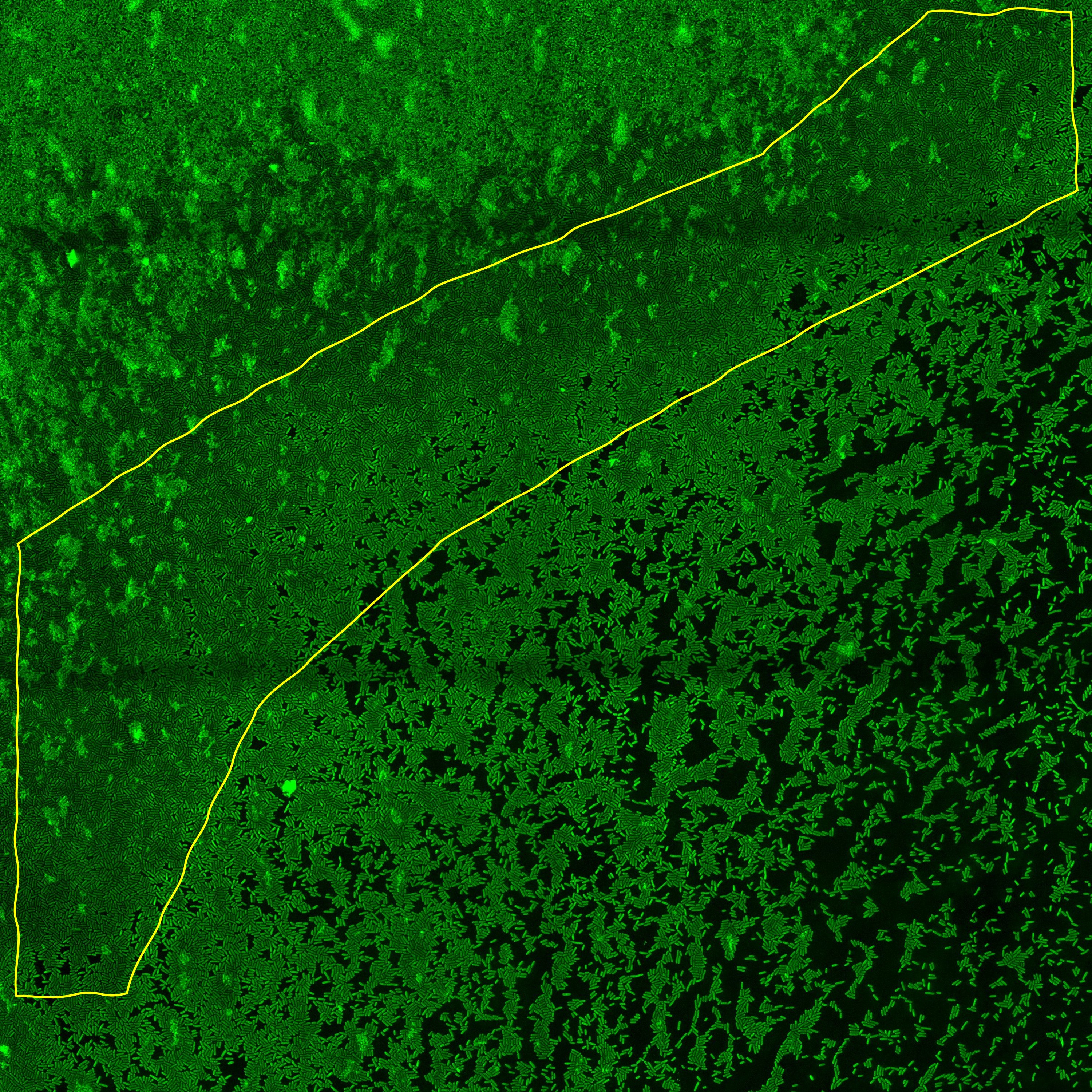
- Look for an area on the slide where bacteria are not diffusing in liquid (while the phages are) and there is a nice monolayer: ideally, every pixel covered by cells, something like the region highlighted in yellow below. This is where you want to record the interactions.
Why agarose pads, and not what the paper describes?
In our 2024 PNAS paper on the microscopic phage adsorption assay, we describe tunnel slides made with double-sided tape, with bacteria stuck to the glass by poly-L-lysine. That is what we used to collect the data in the paper.
That approach works well for E. coli, but it is not as good at keeping other bacteria in place: Pseudomonas, for example, keeps diffusing around, only half-stuck. We have since switched to agarose pads, which hold bacteria tigghtly packed together in place (in a monolayer) while giving them ample nutrients, so they stay metabolically active during imaging.